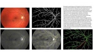
 Our publications for this reporting period have been related to corneal imaging. My colleagues and I have produced methods for the automatic detection of microdots, segmentation of nerves, and building mosaics of images for confocal microscopy. Some of this work can be directly ported to retinal images. Much of the research and hopefully subsequent publications will be related to retinal vessel analysis and image quality. Both are important topics for REVAMMAD. My work on vessel junctions combines local color, edge and gradient information to determine whether the vein or artery crosses on top and analyses the area to determine if knicking is occurring. This work also helps to separate the artery and vein trees in the image.