>
| Roberto Annunziata | University of Dundee |
 |
For full size image: annunziata_progress1 |
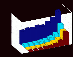
| A novel method for digital curvature estimation has been proposed during this period.A paper has been written and it will be submitted shortly to a journalThis particular area has been investigated since I am working on corneal nerves tortuosity, in collaboration with Harvard Medical School.The problem is to assess image tortuosity and curvature seems to be a good feature for describing tortuosity, even though they are different things. After reviewing the state-of-the-art on digital curvature estimation, I proposed a new solution to unsolved problems in this field: poor accuracy and window size selection.With our multiple-window approach we are now able to estimate digital curvature regardless of the corneal nerve’s size with high accuracy, according to huge amount of experiments on synthetic (ground truth) data. | |
| Carlos Hernandez | FORTH |
 |
For full size image: hernandez_progress1 |
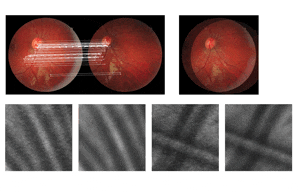
| During this reporting period, I produced an image registration method with sub-pixel accuracy. I employed such method as the first step for already existing super resolution methods, to produce higher quality images from low resolution datasets and submitted a paper. I also started researching on the topic of 3D reconstruction of the eye model employing fundus images as a base. | |
| Georgios Leontidis | University of Lincoln |
 |
For full size image: leontidis_progress1 |
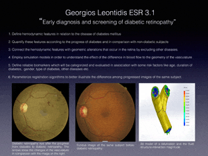
| During this period the main goal was to study the previous and the ongoing research on the early detection of diabetic retinopathy and find all the experiments that have been done until now. At the same time I had to attend a few training events and collaboration meetings at the university of Lincoln with local hospitals as well as internal events. I received some images from the partner in Sunderland which were analyzed for finding some indications of changes in the vasculature before and after diabetic retinopathy. In the meantime, I am trying until now to connect and realize how these geometric changes might be connected with the hemodynamic changes that occur due to diabetes mellitus. Currently I am trying to find more images with specific profile (a few images before and a few after diabetic retinopathy with some information of patients’ history). At the university, we are planning to supply a fundus camera in order to recruit some volunteers for having them screened. This means that we will have images and history record of the volunteers, which will assist on the grouping of the results according to some risk factors like age, gender, duration of diabetes, other diseases etc. | |
| Piotr Chudzik | Orobix SRL |
 |
For full size image: chudzik_progress1 |
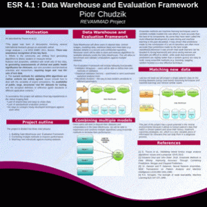
| During last 6 months I reviewed a number of technologies that will be used in implementing the data warehouse and evaluation framework. Together with my supervisor, Dr Luca Antiga, we came up with a very exciting and revolutionary architecture of the data warehouse that will utilize state-of-the-art technologies. We addressed one of the biggest technological challenges of this project, namely how to efficiently store and execute computations alongside data. To accomplish this task we decided to use Docker containers (https://www.docker.io/) which are considered to be “the next big thing” by many experts in the industry that will revolutionize cloud computing. Moreover we came up with a flexible and reproducible way of extracting information from multiple and diverse datasets. | |
| Weiwei Xiang | Charité |
 |
For full size image: xiang_progress1 |
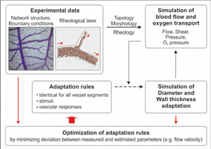
| My REVAMMAD project concerns developing analytical models for simulation of blood flow and vascular structural adaptation in retinal microcirculation under physiological and pathophysiological conditions based on anatomical data from the segmentation of imaging data. In order to gain comprehensive insight into relevant mechanisms underlying function of retinal microvascular network, some other tissues in addition to retina will also be employed for biological experiments. Those extra tissues are expected to supply an easier and comprehensive way for this research because some experimental methods cannot be applied to retina.Significant research during this reporting period:1. Comprehensive theoretical knowledge gained: Insight into morphology, topology, hemodynamics and structural adaptation of microvascular networks which lays theoretical foundation for practical modelling work;2. Hands-on modelling practice in progress: Learning of Delphi programming and apprehension of in-house modelling software on structural adaptation of microvascular network;3. Biological experiments started: Starting to establish the microvascular network occlusion model on CAM. | |
| Sergio Crespo | Charité |
 |
For full size image: crespo_progress1 |
| To approach retinal vascular diseases it is necessary to adopt different strategies to understand the mechanisms of the pathology and transfer that knowledge into practical clinical tools. Vasculature in the retina changes under pathological conditions. However, vessels are not alone and we have to understand the disease as a complex puzzle where every piece has its role. Some other cell types besides the ones that compound vessels might be involved in terms of disease, as it is the case of neurons and microglia. Microglia belongs to the immune system at the retina, but its role in disease it is not clearly understood. Current models of imaging and algorithm analysis are pursuing early diagnosis regarding the dynamics and structure of the vasculature. In our basic medical research, we aim to switch the disease correlation into a new concept that involves vessels inside the neurovascular unit, where microglia might play a role, turning the current analysis model into one more complex and defined status. | |
| Jeff Wigdahl | Uni Padova |
| Most of the work done during this period has been on researching background information for my REVAMMAD project. Side projects have included work in confocal microscopy related to the detection of features in different layers of the cornea. This work has led to the submission of two abstracts, one of which has already been accepted and will be presented as a poster at the ARVO conference. The other was submitted to the EMBC conference. Corneal nerves are similar to retinal vessels and many of the nerves metrics can also be calculated for vessels, such as tortuosity. This overlap will lead to much of the work being produced now, to be useful when the Retinal vessel work is undertaken. | |
| Giovanni Ometto | Aarhus |
 |
For full size image, ometto-progress-1 |
| NUMBER AND LOCATION OF LESIONS FOR THE OPTIMIZATION OF THE EXAMINATION INTERVAL IN DIABETIC RETINOPATHYA recent study concluded that it is possible to construct a model for optimising the examination interval during screening for diabetic retinopathy in low-‐risk patients. However, the model fails to predict the interval for patients in whom the primary assessment recommended a short screening interval, suggesting that more risk factors should be identified and included. Two sets of fundus photographs, one where the result of the model and recommended interval are concordant and one where they are discordant, were analysed. The number of different lesions and their centre position (see fig. 1) with regards to the areas defined on a previous study (see fig. 2) were stored in an array of features associated with the relative fundus photo (see table 1). Considering the concordance and discordance on the interval as the two possible values of a dependent variable in a classification problem, an extraction of the best isolated features and a 10-‐fold cross validation were performed to assess their significance. The feature selection chose as the two best isolated features the number and the percentage of small haemorrhages in two different areas respectively. The 10-‐fold cross validation performed extracting the first two ranked features only resulted in the correct classification of 64% of the instances. With these two sets the ranking of the feature selection matched the clinicians’ experience while the percentage of correct classifications suggested that the model would benefit from the information held in the location and number of small haemorrhages showing on fundus photography. | |
| Pedro Guimarães | Uni Padova |
 |
For full size image,guimaraes-arvo-poster |
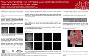
| During this reporting period, the first version of the algorithm to segment corneal endothelial cells was developed. The corneal nerves segmentation algorithm was adapted and tested in mosaic images. | |
| Matteo Aletti | INRIA |
 |
For full size image, aletti_progress1 |
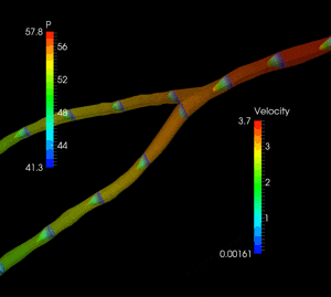
| In the first six month working on the REVAMMAD project we developed an automatic algorithm to generate a 3D mesh of the retinal vessels starting from segmented images of retinal fundus from Lincoln University. We also devised an ad hoc fluid structure interaction model for modeling the interaction between blood and vessel wall into the retinal vasculature. We included in the model for the vessel wall the regulation of the blood flow rate. We are currently summarizing the results in view of their publication. | |