Evi Kotsiliti – Progress Report 3

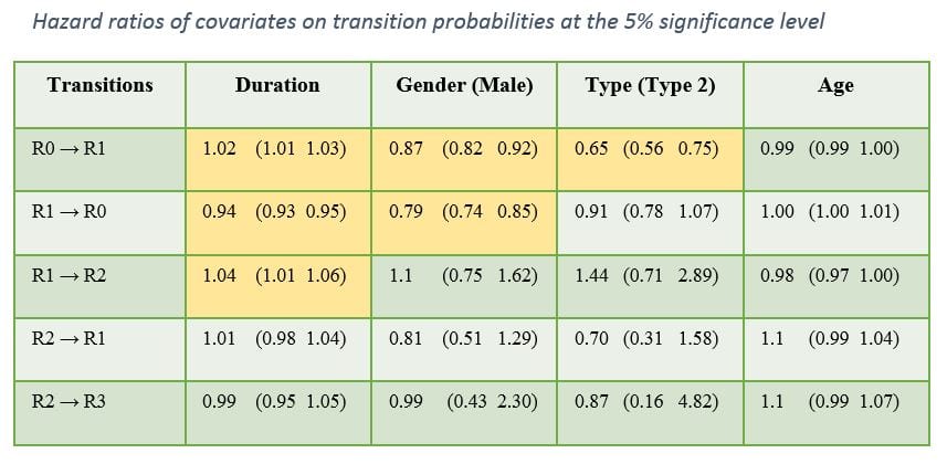
In this reporting period, the risk of progression to the different states of diabetic retinopathy (R0, R1, R2 and R3) was estimated based on data collected from patients attending systematic retinal screening in the region of East Anglia. There was a total of 14,348 patients who were followed up for a time period of 6 years for whom the duration of diabetes, type of diabetes, age and gender were recorded.

The risk could be estimated for the cohort as well as for each individual based on their clinical profile. During the validation stage, the observed number of patients in each state was compared against the expected number of patients according to the risk estimation. There was a good approximation for the first two states and a mediocre approximation for the last two, primarily due to a small number of available data for these two states. The duration of diabetes, the type of diabetes and the gender were found to have a significant effect on the risk of transitioning in some states as can be seen in the picture attached.

For future steps, I wish to enhance the accuracy of the risk estimation by deploying more data from the more advanced states and more predictors.

Picture_reporting period march - september 2015


Jeff Wigdahl – Progress Report 4

This reporting period was mostly about collaborations. I was able to work with many of the other ESRs and co-author a few conference papers with them. It also gave me the opportunity to collect some data from the University of Dundee to help create a new tortuosity testing dataset. Once available, the dataset will allow others to publicly download and test new metrics while comparing to multiple clinician gradings. I was also able to create and publish my method for detecting vessels crossover abnormalities. This adds another tool to the retinal vessel analyser, the main topic of my project.

Weiwei Xiang – Progress Report 4

During this period of research, the following was achieved:

  • Finalization of experiment protocol on adaptive remodelling of microvascular networks in CAM after acute microocclusion in arterioles and venules.
  • In collaboration with ESR 2.4, Febro Guimaraes: Semi-automatic velocity measurement software tested.
  • In collatoraction with ESR 2.1, Francesco Caliva, and Dr Bashir Al-Diri: Preliminary test on network reconstruction of CAM microvasculature.


Carlos Hernandez Matas – Progress Report 4

Carlos Hernandez Matas Progress report 4 slide

During this research period, Carlos improved his retinal image registration method, which aligns retinal images. This is useful for studying the evolution of disease across examinations, or to enable other techniques such as the creation of super resolution images to perform more accurate measurements or study fluid dynamics on the retinal vessels when the images are taken in the same session.

Anastasiia Neganova – Progress report 4

BMIday2015 dec

During this reporting period, the model of the retinal vascular network was improved and myogenic response gradient through the network was implemented. Major results for a modelling paper were obtained. A draft of paper on modelling was written.

Also, animal experiments were carried out: set-up and methods were improved. Experiments focused on retinal reaction on chemicals of normal rats. Vessels in inner retina were successfully detected and raw data now is analysed. In the near future, writing of a draft on results of experiments is planned.

Two months were spent in Berlin, where the secondment took place. There, proper working with egg’s embryos were studied. LSI techniques in order to measure blood flow in CAM during vascular remodelling were applied.

Matteo Aletti – Progress Report 4

These six months were mainly dedicated to the dissemination of my work: in fact, we have submitted two publications and I presented the results in two different conferences: USNCCM about computational mechanics and CMBE more related to bioengineering. At the same time we have continued working on the reduced order modeling techniques to be used in the modeling of the Intra Ocular Pressure.

Giovanni Ometto – Progress Report 4

Photo - progress report 4The existing lesion-detection algorithm was improved with the introduction of a new template-detection method. The resulting hybrid algorithm showed higher sensitivity than the previous one. A new method for the quick detection of the optic disk was developed. A new method for the fully automatic detection of 5 clinically relevant regions was developed.

Francesco Caliva – Progress Report 4

Francesco report October 2015

During this reporting period, the development of a tool usable to connect flood vessels in fundus images gave a contribution to the retinal community. This work, which was published at IEEE EMBC 2015, presented a tool that used implicit neural cost functions to join retinal vessels. Results obtained showed that the quality of the segmentation affected the outcome of connectivity algorithms and by enhancing the segmentation the connectivity was improved. Research focused also on arteries and veins recognition in fundus image. This is a topic of high interest in order to insight the study of diseases. An algorithm able to classify arteries and veins after segmenting vessels is currently being implemented and tested.

Collaboration was carried out with ESR 3.3 Giovanni Ometto. In this work, a fast method for the definition of clinically relevant retinal areas in use for screening programmes for diabetic retinopaghy was produced.

Roberto Annunziata – Progress Report 4

qualitative_comparison Oct 15

The fully automated framework for curvilinear structure tortuosity quantification has been completed and tested extensively. It consists of the combination of a new ridge detector designed and developed for detecting tortuous structures (SCIRD) and learned context filters. Qualitative (the image attached shows a comparison of our curvilinear structur detector, SCIRD and state-of-the-art methods) and quantitative results show that our system compare favourably with previously reported methods and achieve a level of accuracy comparable or better than three experience observers. The original building blocks of the proposed framework have been accepted as three MICCAI conference papers and a journal paper presenting the system will be submitted soon. Please refer to my webpage for further details.

Sergio Crespo Garcia – Progress Report 4

IMG_8352Microglia play a major role in retinal neovascularization and degeneration and are thus potential targets for therapeutic intervention. In vivo assessment of microglia behaviour in disease models can provide important information to understand patho-mechanisms and develop therapeutic strategies. Although scanning laser ophthalmoscope (SLO) permits the monitoring of microglia in transgenic mice with microglia-specific GFP expression, there are fundamental limitations in reliable identification and quantification of activated cells. Therefore, we aimed to improve the SLO-based analysis of microglia using enhanced image processing with subsequent testing in laser-induced neovascularization (CNV). CNV was induced by argon laser in MacGreen mice. Microglia was visualized in vivo by SLO in the fundus auto-fluorescence (FAF) mode and verified ex vivo using retinal preparations. Three image processing algorithms based on different analysis of sequences of images were tested. The amount of recorded frames was limiting the effectiveness of the different algorithms. Best results from short recordings were obtained with a pixel averaging altorithm, further used to quantify spatial and temporal distribution of activated microglia in CNV. Morphologically, different microglia populations were detected in the inner and outer retinal layers In CNV, the peak of microglia activation occurred in the inner layer at day 4 after laser, lacking an acute reaction. Besides, the spatial distribution of the activation changed by the time over the inner retina. No significant time and spatial changes were observed in the outer layer. An increase in laser power did not increase number of activated microglia. The SLO, in conjuction with enhanced image processing, is suitable for in vivo quantification of microglia activation. This surprisingly revealed that laser damage at the outer retina led to more reactive microglia in the inner retina, shedding light upon a new perspective to approach the immune response in the retina in vivo.

Georgios Leontidis – Progress Report 4

Georgios_progress_4During this period, I focused mainly on extracting as many features as possible from the available DR images based on strict quality criteria (regarding inclusion of observations, power estimations and avoidance of statistical errors). Moreover, a suitable hybrid mixed model was introduced specific for the nature of my analysis which can be used when the independence of observations is violated in simple models where we include correlated data. The fractality and lacunarity for the complexity of vascular trees has been introduced for which the skeletonised or segmented images are required. Multiple features are being tested for their significance and discriminative ability identifying patterns of changes when the vessels’ structrure is changing. New features include branch retinal equivalent, width-to-angle ratio, sum of squares of widths (which is continuously differentiable and useful for minimising variance) and can be used in the cross section estimation which in turn is useful for the new flow rate function. Centreline length to straight distance ratio and the junction exponent calculated by the Branching coefficient and fractal dimension are among the other possible features. CRVE/CRAE-AVR and tortuosity have been incorporated into the analysis.