ESRs research progress

Anastasiia Neganova – Progress report 4

BMIday2015 dec

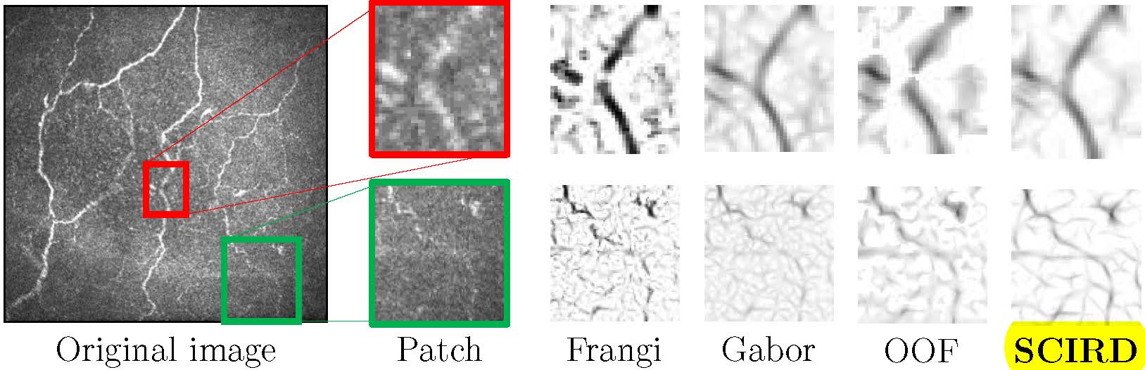
During this reporting period, the model of the retinal vascular network was improved and myogenic response gradient through the network was implemented. Major results for a modelling paper were obtained. A draft of paper on modelling was written.

Also, animal experiments were carried out: set-up and methods were improved. Experiments focused on retinal reaction on chemicals of normal rats. Vessels in inner retina were successfully detected and raw data now is analysed. In the near future, writing of a draft on results of experiments is planned.

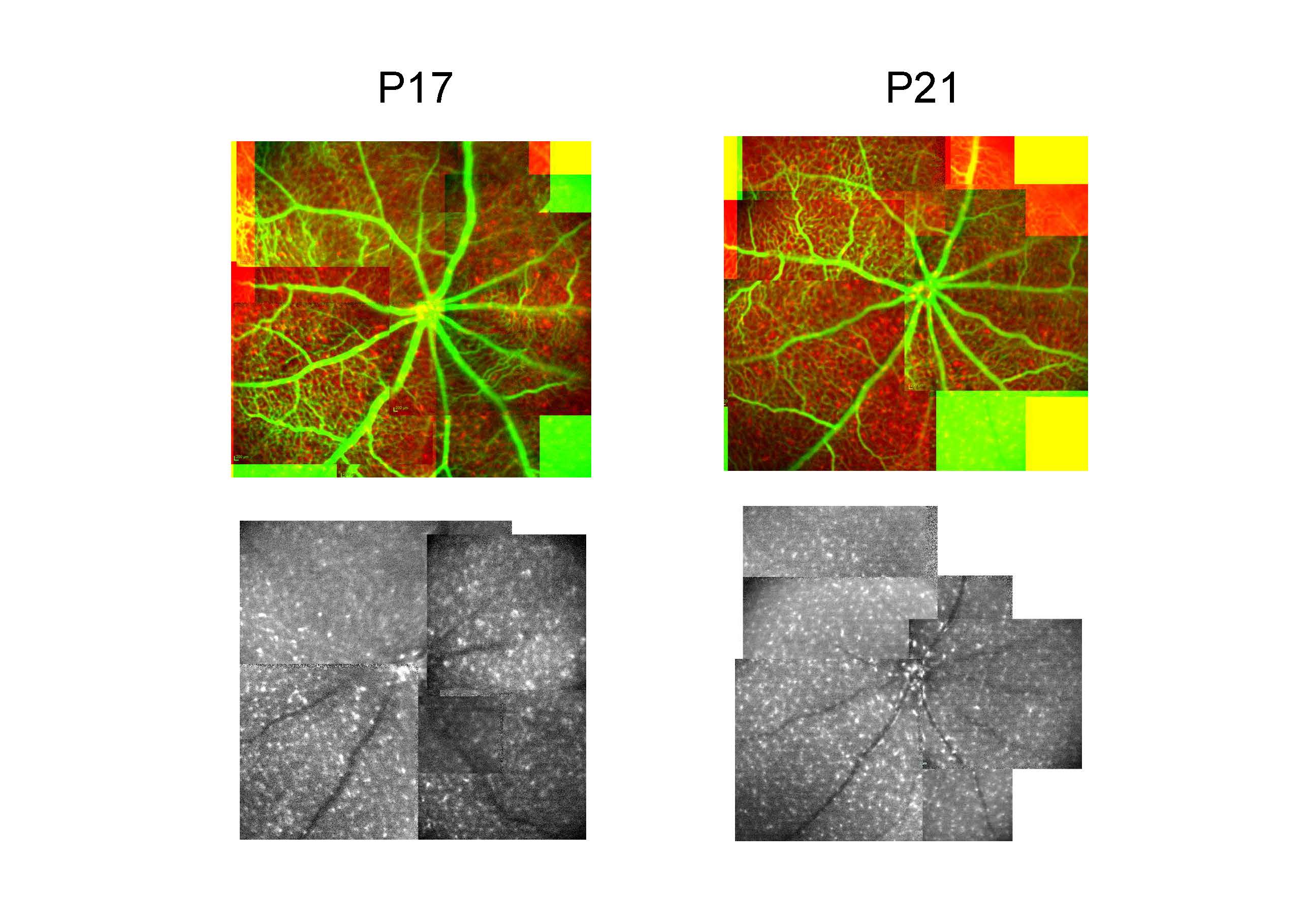
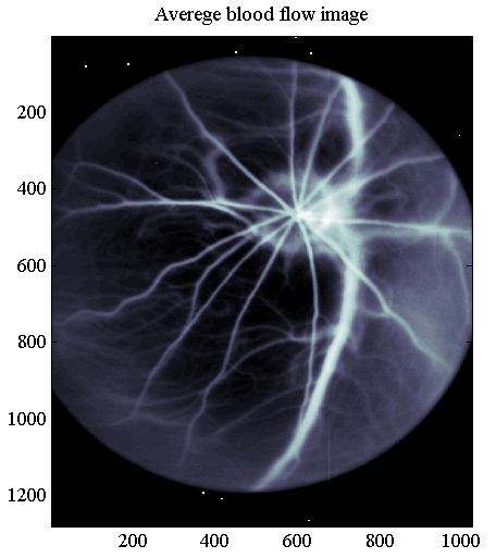
Two months were spent in Berlin, where the secondment took place. There, proper working with egg’s embryos were studied. LSI techniques in order to measure blood flow in CAM during vascular remodelling were applied.

Matteo Aletti – Progress Report 4

These six months were mainly dedicated to the dissemination of my work: in fact, we have submitted two publications and I presented the results in two different conferences: USNCCM about computational mechanics and CMBE more related to bioengineering. At the same time we have continued working on the reduced order modeling techniques to be used in the modeling of the Intra Ocular Pressure.

Giovanni Ometto – Progress Report 4

Photo - progress report 4The existing lesion-detection algorithm was improved with the introduction of a new template-detection method. The resulting hybrid algorithm showed higher sensitivity than the previous one. A new method for the quick detection of the optic disk was developed. A new method for the fully automatic detection of 5 clinically relevant regions was developed.

Francesco Caliva – Progress Report 4

Francesco report October 2015

During this reporting period, the development of a tool usable to connect flood vessels in fundus images gave a contribution to the retinal community. This work, which was published at IEEE EMBC 2015, presented a tool that used implicit neural cost functions to join retinal vessels. Results obtained showed that the quality of the segmentation affected the outcome of connectivity algorithms and by enhancing the segmentation the connectivity was improved. Research focused also on arteries and veins recognition in fundus image. This is a topic of high interest in order to insight the study of diseases. An algorithm able to classify arteries and veins after segmenting vessels is currently being implemented and tested.

Collaboration was carried out with ESR 3.3 Giovanni Ometto. In this work, a fast method for the definition of clinically relevant retinal areas in use for screening programmes for diabetic retinopaghy was produced.

Roberto Annunziata – Progress Report 4

qualitative_comparison Oct 15

The fully automated framework for curvilinear structure tortuosity quantification has been completed and tested extensively. It consists of the combination of a new ridge detector designed and developed for detecting tortuous structures (SCIRD) and learned context filters. Qualitative (the image attached shows a comparison of our curvilinear structur detector, SCIRD and state-of-the-art methods) and quantitative results show that our system compare favourably with previously reported methods and achieve a level of accuracy comparable or better than three experience observers. The original building blocks of the proposed framework have been accepted as three MICCAI conference papers and a journal paper presenting the system will be submitted soon. Please refer to my webpage for further details.

Sergio Crespo Garcia – Progress Report 4

IMG_8352Microglia play a major role in retinal neovascularization and degeneration and are thus potential targets for therapeutic intervention. In vivo assessment of microglia behaviour in disease models can provide important information to understand patho-mechanisms and develop therapeutic strategies. Although scanning laser ophthalmoscope (SLO) permits the monitoring of microglia in transgenic mice with microglia-specific GFP expression, there are fundamental limitations in reliable identification and quantification of activated cells. Therefore, we aimed to improve the SLO-based analysis of microglia using enhanced image processing with subsequent testing in laser-induced neovascularization (CNV). CNV was induced by argon laser in MacGreen mice. Microglia was visualized in vivo by SLO in the fundus auto-fluorescence (FAF) mode and verified ex vivo using retinal preparations. Three image processing algorithms based on different analysis of sequences of images were tested. The amount of recorded frames was limiting the effectiveness of the different algorithms. Best results from short recordings were obtained with a pixel averaging altorithm, further used to quantify spatial and temporal distribution of activated microglia in CNV. Morphologically, different microglia populations were detected in the inner and outer retinal layers In CNV, the peak of microglia activation occurred in the inner layer at day 4 after laser, lacking an acute reaction. Besides, the spatial distribution of the activation changed by the time over the inner retina. No significant time and spatial changes were observed in the outer layer. An increase in laser power did not increase number of activated microglia. The SLO, in conjuction with enhanced image processing, is suitable for in vivo quantification of microglia activation. This surprisingly revealed that laser damage at the outer retina led to more reactive microglia in the inner retina, shedding light upon a new perspective to approach the immune response in the retina in vivo.

Georgios Leontidis – Progress Report 4

Georgios_progress_4During this period, I focused mainly on extracting as many features as possible from the available DR images based on strict quality criteria (regarding inclusion of observations, power estimations and avoidance of statistical errors). Moreover, a suitable hybrid mixed model was introduced specific for the nature of my analysis which can be used when the independence of observations is violated in simple models where we include correlated data. The fractality and lacunarity for the complexity of vascular trees has been introduced for which the skeletonised or segmented images are required. Multiple features are being tested for their significance and discriminative ability identifying patterns of changes when the vessels’ structrure is changing. New features include branch retinal equivalent, width-to-angle ratio, sum of squares of widths (which is continuously differentiable and useful for minimising variance) and can be used in the cross section estimation which in turn is useful for the new flow rate function. Centreline length to straight distance ratio and the junction exponent calculated by the Branching coefficient and fractal dimension are among the other possible features. CRVE/CRAE-AVR and tortuosity have been incorporated into the analysis.


Jeff Wigdahl – Progress Report 3

Our publications for this reporting period have been related to corneal imaging. My colleagues and I have produced methods for the automatic detection of microdots, segmentation of nerves, and building mosaics of images for confocal microscopy. Some of this work can be directly ported to retinal images. Much of the research and hopefully subsequent publications will be related to retinal vessel analysis and image quality. Both are important topics for REVAMMAD. My work on vessel junctions combines local color, edge and gradient information to determine whether the vein or artery crosses on top and analyses the area to determine if knicking is occuring. This work also helps to separate the artery and vein trees in the image.

Matteo Aletti – Progress Report 3

In the last nine months I have been mainly working on three different topics: smooth muscle models, retinal image analysis and intra ocular pressure (IOP). These topics may seem unrelated, however, retinal imaging tools are fundamental in providing reliable geometries for haemodynamics models. Retinal blood flow is characterized by autoregulation phenomena and smooth muscle cells play an important role in it: contracting and relaxing following bio-chemical stimuli. We have summarized the results of this autoregulation models in a publication that is going through the review process. Finally, IOP plays a major role in several eye diseases (glaucoma, for instance) and we have just started to address the problem of modeling of the eye from a broader point of view coupling the haemodynamics with the intra ocular pressure.


Evi Kotsiliti – Progress Report 2

PGR conference_posterThe main goal of my project is to define alternative strategies that can prove to be more efficient and/or cost saving in the detection of diabetic retinopathy. The first step of this process was an extensive literature review in order to gain a comprehensive understanding of the nature of the disease, the risk factors that affect its progression and the different frameworks applied for detection. My knowledge was further reinforced by untertaking the level 3 City and Guilds accreditation for screeners and graders and by participating in respective conferences and seminars.

Currently, I am trying to define the underlying parameters of the National Diabetic Eye Screening Programme and estimate the relative costs especially in the case of inappropriate decisions. This is the starting point of my practical implementation and the accurate definition of the framework of this model is crucial in the further assessment of alternative approaches.

Carlos Hernandez Matas – Progress Report 3

During this reporting period, Carlos continued working on his 3D retinal registration method, obtaining very promising experimental results. Those results lead to the preparation of a paper that will be submitted to Engineering in Medicine and Biology Conference 14. He also worked with ESR 3.2, Sergio Crespo Carcia, in a fruiful collaboration that led to the submission of a paper to the Experimental Eye Research Journal.


Weiwei Xiang – Progress Report 3

Image from WeiweiMy work over the past six months falls into three categories:

  1. Establishment of protocols on wall thickness measurement of microvessels (diameter>=10 µm) on chick chorioallantoic membrane (CAM)
  2. Devising of protocols on videomicroscopy using high speed camera which allows pulsatility analysisi of blood flow on CAM
  3. Semi-automatic segmentation methods established in collaboration with ESR 2.1 Francesco Caliva and Dr. Bashir Al-Diri from University of Lincoln, UK.

Giovanni Ometto – Progress Report 3

picture 3

The automatic detection of lesions is a fundamental tool for my future research on the progression of diabetic retinopathy. My secondment in Lincoln was aimed at testing and adjusting an existing algorithm available at my partner University. While the algorithm was created to find all types of lesion to automate the diagnosis of referable maculopathy, my future research will need it for the detection of micro-aneurisms.

Testing the algorithm on the pictures from high-risk patients from my previous study showed that more than half of micro-aneurisms were not detected. It was found that many of the lesions were not detected on the candidate-selection stage of the algorithm. A newly developed method, based on the template matching technique, could find 50% more true-lesions candidates.

The combination of the two methods detects approximately 70% of the true lesions among the candidates from funds photographs from high-risk patients.

Sergio Crespo Garcia – Progress Report 3

I am participating actively in several projects that approach neovascularization under pathological conditions in the retina, with the interaction of an inflammatory or hypoxic environment. Projects to mention are:

  • Quantification of activated microglia in vivo in laser CNV MacGreen mouse.
  • Settling of a model for hypertensive retinopathy: dTGR.
  • Settling of a model for diabetic retinopathy in a knock down rat: TetO.
  • Analysis in vivo and in vitro of CD11b+ cells under ROP paradigm. .
  • The role of Netrin4 under pathological neovascularization.
  • In vivo follow-up of macrophages in early stages of STZ MacGreen mouse and comparison with a double model MacGreen-Netrin4-/-.
  • Role of PlGF in pathological neovascularization.
  • Chronic bilateral cranial hypoperfusion in mice analysis in the retina.

figure

Roberto Annunziata – Progress Report 3

I have been working on the design and development of ad-hoc methods for curvilinear structures segmentation.

This work was motivated by the fact that state-of-the-art approaches tend to fail when dealing with corneal nerve fibres captured via confocal microscopy. Qualitative and quantitative results show that our methods outperform previously reported methods on several dataset.

qualitative_results

Automated curvilinear structure detection in images capturing corneal nerve fibres and neuronal trees with various technical challenges such as confounding non-target structures, low contrast, low resolution, non-uniform illumination, high tortuosity level and fragmentation.

Georgios Leontidis – Progress Report 3

Georgios report 3During this period, I focused mainly on extracting as many features as possible from the available DR images, based on strict quality criteria (regarding inclusion of observations, power estimations and avoidance of statistical errors). Moreover, a suitable hybrid mixed model was introduced specific for the nature of my analysis which can be used when the independence of observations is violated in simple models where we include correlated data.

The fractality and lacunarity for the complexity of vascular trees has been introduced for which the skeletonised or segmented images are required. Multiple features are being tested for their significance and discriminative ability identifying patterns of changes when the vessels’ structure is changing.

New features include branch retinal equivalent, width-to-angle ratio, sum of squares of widths (which is continuosly differentiable and useful for minimising variance) and can be used in the cross section estimation which in turn is useful for the flow rate function. Centreline length to straight distance ration and the junction exponent calculated by the Branching coefficient and fractal (box-counting/hausdorf) dimention are among the other possible features.

Francesco Caliva – Progress Report 3

MATLAB Handle GraphicsDue to image artefacts, the result of the blood vessels’ segmentation is a set of disconnected short segments. During this three months reporting period, I worked on the problem of connecting them. At this aim, an algorithm was implemented.

Given a group consisting of a certain number of segments, there can be connected together in several ways: two segments can join each other and form a bridge; three segments can generate a bifurcation; or they can just be terminals.

At this stage, the connections rely on the use of implicit cost functions.

Besides, an user friendly Matlab tool was designed. Thanks to this tool, it is possible to adjust the results obtained using the previously mentioned cost functions. The tool includes also a fast altorithm for the segmentation of missing blood vessels.

The tool, which in part was developed with ESR 1.1 during his secondment at the University of Lincoln, is currently being tested by ESR 1.2.


Anastasiia Neganova – Progress Report 3

RetinaRat2MeanDuring this reporting period, the mathematical model of the retinal vascular network was improved, and some results for paper were obtained. Work on manuscript, dedicated to mathematical model was started. In addition to modeling part of my work, animal experiments on rats eyes were carried out. Now, these experiments are focused on detection of retinal reaction on flickering-light stimulus. Furthermore, I am involved in experiments on eggs chorioallantoic membrane, which are focused on vascular network remodelling. To get the rat retinal images and images of eggs chorioallantoic membrane we using Laser Speckle Contrast method, that is “full-field” and non invasive technique.

Weiwei Xiang – Progress Report 2

2nd illustrationExperimentation, as part of my REVAMMAD project, aims to lay ground for simulation of blood flow and vascular structural adaptation in microvascular network on CAM before and after arteriolar occlusion. Generally, this experimental work includes three consecutive components: 1) Cultivation of ex-ovo chicken embryos; 2) Imaging and recording of microvascular network on ex-ovo CAM assay with and without laser irradiation; 3) Parameter analysis including parameters of vessel morphology, network topology, hemodynamics, metabolism and vascular adaptation.

Sergio Crespo Garcia – Progress Report 2

ProgressReport2ImageTo approach retinal vascular diseases it is necessary to adopt different strategies to understand the mechanisms of the pathology and transfer that knowledge into practical clinical tools. Vasculature in the retina changes under pathological conditions. However, vessels are not alone and we have to understand the disease as a complex puzzle where every piece has its role. Some other cell types besides the ones that compound vessels might be involved in terms of disease, as it is the case of neurons and microglia. Microglia belongs to the immune system at the retina, but its role in disease it is not clearly understood. Current models of imaging and algorithm analysis are pursuing early diagnosis regarding the dynamics and structure of the vasculature. In our basic medical research, we aim to switch the disease correlation into a new concept that involves vessels inside the neurovascular unit, where microglia might play a role, turning the current analysis model into one more complex and defined status.

ProgressReportFigure

Roberto Annunziata – Progress Report 2

progress_report_2_repimgClassify in vivo confocal microscopy corneal images by tortuosity is complicated by the presence of variable numbers of fibres of different tortuosity level. Instead of designing a function combining manually selected features into a single coefficient, as done in the literature, we proposed a supervised approach which selects automatically the most relevant combination of shape features from a pre-dened dictionary. To our best knowledge, we are the first to consider features at different spatial scales and show experimentally their relevance in tortuosity modelling. Experimental results using a data set of 90 images provided by our clinical collaborators at the Harvard Medical School and Massachusetts Eye and Ear Infirmary show that our framework yields an accuracy indistinguishable, overall, from that of experts when compared against each other.

Piotr Chudzik – Progress Report 2

piotr_chudzik_progress_report_slideDuring last 6 months I managed to accomplish an important milestone in my project, namely the creation of a prototype of Data Warehouse. Initial architecture decisions proved to be correct and led to an implementation that is robust and flexible at the same time. Existence of a prototype  allows me to start incorporating datasets and computations into the system as well as commence testing of all components interacting with each other. Data warehouse is built using innovative technologies and we believe that it has a strong industrial potential because (to the best of our knowledge)  there is no similar solution in the market. The existence of  Data Warehouse which stores multiple heterogeneous datasets and computations that makes it easy to query them is essential before proceeding to the next part of the project which is extracting similar retinal images. After reviewing scientific literature dealing with similarity learning I decided to use deep learning which excels at learning  intermediate representations of highly dimensional data that can be used to learn similarity between medical images.  My deep learning research led me to a discovery of a novel approach for retinal vessel segmentation that can compete with the state-of-the-art methods in this field.

Jeff Wigdahl – Progress Report 2

ProgressReport2JWOur publications for this reporting period have been related to corneal imaging.  My colleagues and I have produced methods for the automatic detection of microdots, segmentation of nerves, and building mosaics of images for confocal microscopy.  Some of this work can be directly ported to retinal images.  Much of the research and hopefully subsequent publications will be related to retinal vessel analysis and image quality.  Both are important topics for REVAMMAD.  My work on vessel junctions combines local color, edge and gradient information to determine whether the vein or artery crosses on top and analyses the area to determine if knicking is occurring.  This work also helps to separate the artery and vein trees in the image.

Giovanni Ometto – Progress Report 2

Picture2 Ometto Picture1 Ometto

Distribution of miicroanreurisms of the dataset

Number and location of lesions for the optimisation of the screening interval in diabetic retinopathy

Diabetic retinopathy is a leading cause of blindness in the Western World. The disease is detected by screening which includes visual acuity measurement and fundus photography (Stefansson et al. 2000; Jeppesen & Bek 2004; WHO 2008; Bandello et al 2013). On the basis of the diabetes type and the severity of retinopathy a fixed interval to the following screening examination defined to ensure that no patient will develop vision threatening complications during that period (Singer et al. 1992; AAO Diabetic Retinopathy PPP 2012). However, this rule-based approach also implies that patients with slow disease progression will experience a number of superfluous examinations without consequences for the management of the disease. A reduction in the number of these examinations requires an algorithm that considers the patient’s individual risk factors (Aspelund et al 2011). The inclusion of risk factors such as sex, age at onset of diabetes mellitus, diabetes type, diabetes duration, HbA1c and blood pressure has been shown to allow a significant prolongation of the control interval without increasing the risk of developing vision threatening retinopathy (Mehlsen et al. 2012). The predictive value of this model was found to be lowest for patients with more severe retinopathy, and it was concluded that the grader might have included other factors than the severity of retinopathy in the decision, such as the location of retinopathy lesions or conditions unrelated to diabetic retinopathy (Hove et al. 2004, 2006). Therefore, 81 patients out of 6868 observations where the prediction of the model matched exactly or deviated significantly (3 or more time intervals) from the clinician’s recommended interval were identified. The fundus photographs documented during the screening visits from these patients were reviewed in order to investigate the role of the location of retinopathy lesions and the role of the presence of retinal lesions unrelated to diabetic retinopathy in the assessment of control intervals of the same or different lengths in time, whether assessed using the rule based approach or the individualised model.


Georgios Leontidis – Progress Report 2

presentation_leontidis_annualreportDuring this period, the main goal was to go deeper in the analysis of diabetic patients’ images and try to find any possible indications of the early changes of the vasculature. Moreover the writing of a comprehensive review paper helped to go deeper in understanding the underlying mechanisms that affect the auto regulation of the retina. Many progressed images of patients were analyzed by measuring multiple matched segments both for veins and arteries, mainly focusing on the widths and branching angles as well as the location of the vessels. This research gave me the opportunity to present a poster with my research in a medical imagining school and publish the results at an international conference, getting the feedback from other experts.