Matteo Aletti – Progress Report 6

During the last 9 months I have mainly worked on the development of a full model of the eye. In particular I have developed a poro-elastic model for the choroid and I have coupled it with a simple model of the vitreous. To obtain these results I have worked on the development of a library that can be used to couple different solvers. The idea is to develop adhoc software for the simulation of each compartment of the eye (for example: the choroid, the vitreous, the cornea, the anterior chamber, etc…) and make them interact with each other by using this library.

I have also finalised the publication on the reduced order technique that we have developed to speed up those simulations, alongside writing my PhD thesis.

Georgios Leontidis – Progress Report 6

In the final 6 months of the project I mainly focused on two things: Firstly, I closely collaborated with another ESR in order to apply mathematical modelling in estimating haemodynamic features. This was applied in the context of identifying important biomarkers of progression to diabetic retinopathy. Our common work concluded with writing up a journal paper, which was submitted and published. Secondly, I also focused on writing up my PhD thesis, recapping on all of the research that I conducted during my MSCA fellowship. The thesis was submitted at the end of my contract and I successfully passed my examination.

Evi Kotsiliti – Progress Report 6

In the last reporting period, a model for predicting patients at risk of developing diabetic retinopathy was designed based on patient characteristics and clinical measurements. The model yielded a cross-validated AUC of 0.77 ± 0.04 and could be used to identify patients at risk of developing retinopathy, resulting to a significant reduction of the number of screening visits.

The below poster was also presented at the European Researchers’ LiGHTS Nights 2016 Science Festival hosted by the University of Lincoln:

poster_EU research nights lights 2016_v2

Jeff Wigdahl – Progress Report 6

This reporting period my main contribution has been my work in optic disc detection and curvature enhancement. Both methods Progress Report 6proposed were able to outperform state of the art methods on publicly available datasets. Both methods are also very adaptable (this being the main feature of the tortuosity estimation). The curvature enhancement method has allowed for a new set of tortuosity metrics to be created. By changing the filter banks, these metrics can be adapted to better estimate the tortuosity evaluation of an individual grader.


Figure 1: Examples of different curvature enhancements on a set of synthetic vessels using the proposed method for curvature enhancement.

Francesco Caliva – Progress Report 6

The progression from diabetes to diabetic retinopathy is associated with changes in retinal haemodynamics. In collaboration Progress Report 6with Georgios, a longitudinal study of twenty-four subjects was conducted. In this study, we monitored the retinal haemodynamics during the three years before the appearance of diabetic retinopathy (DR) and in the first year of DR. We took vascular measurements from standard fundus images, and estimated fluidynamic parameters using a simple haemodynamic model. We show that there are statistically significant changes in some estimated haemodynamic parameters associated with the development of DR.
Please refer to the related paper “Hemodynamics in the retinal vasculature during the progression of diabetic retinopathy” (F. Calivá, G. Leontidis, P. Chudzik, A. Hunter, L. Antiga, B. Al-Diri), which was successfully submitted to the Journal for modeling in Ophthalmology.

Roberto Annunziata – Progress Report 6

During the last period, I have been focusing on revising our Medical Image Analysis paper presenting and validating the whole framework for automated corneal nerve image tortuosity estimation and interpretation. Moreover, I have been drafting and revising an IEEE Transactions on Medical Imaging paper on accelerating convolutional sparse coding for curvilienar structure segmentation and a new, more robust version of the SCIRD filters (see Figure) I proposed at MICCAI 2015. In the last part of the period I have been writing my PhD thesis and successfully passed the viva-voce examination on the 12th of Aug, subject to some very minor revisions. In parallel, I have tested the system I developed on a large data set of ~500 images from subject with different pathologies and a clinical paper is being written by our collaborators at Harvad Medical Schiool and Tufts Medical Center to report the main outcomes.
For more information, please go to: http://staff.computing.dundee.ac.uk/rannunziata

Progress Report 6 - Slides (DHB)

Weiwei Xiang – Progress Report 6

The final results of my project show that after occlusion, unidirectional flow towards the occlusion site was established. Diameters exhibited an immediate decrease, an ensuing increase with a time constant of 2.53 ± 0.77h (mean ± SD, n = 13 CAMs) and then a plateau of up to 60% above baseline without significant vascular tone change. WSS exhibited an immediate increase, a linear decrease to baseline approximately 12h post-occlusion and then remained unchanged. WSS increase upon occlusion and sustained diameter changes were correlated. WSS change only partly mirrored the diameter change rate through 24h post-occlusion.

Giovanni Ometto – Progress Report 6

The algorithm for the automatic detection of the ONH is improved and is now the fastest and second best (0.26% difference) in terms of performance compared to the best in literature, when tested on MESSIDOR dataset. Qualitative results are also improved with an elliptical approximation of the ONH boundary. This was possible by describing the photograph distortion is caused by the quasi-spherical shape of the eye.

A probability map is created based on a prospective database. The database includes 60 patients: 30 developed vision threatening DR / 30 did not. Approximately 10-year record from screening database for each patient (for an average of 7 visits each). The dataset includes more than 900 images and the analyses is made possible with the tools previously developed. The map returns the probability of developing vision threatening diabetic retinopathy given the location of the first detected microaneurysm for patients included in a diabetic retinopathy screening programme.

Example result from the ONH detection with elliptical correction:

Progress Report 6 image1


The model of the eye used to quantify the distortion due to the orthogonal projection
of a curved surface:

Progress Report 6 image2


Probability of developing vision threatening diabetic retinopathy given the location of
the first detected microaneurysm for patients included in a diabetic retinopathy
screening programme:

Progress Report 6 image3

Carlos Hernandez Matas – Progress Report 5

During this reporting period, Carlos conducted research regarding the utilisation of multi-modal cues as well as the accuracy of eye shape estimation for the retinal image registration method. Additionally, an image dataset for retinal image registration (FIRE: Fundus Image Registration Dataset) was made publicly available at http://www.ics.forth.gr/cvrl/fire/ containing 134 image pairs falling under 3 different categories and including ground truth for registration evaluation.Slide1

Sergio Crespo García – Progress Report 6

ProgressReport6_figureType 2 diabetes represents the most prevalent cause for progression of diabetic retinopathy. However, there is a lack of appropriate translation animal models. Although the renin-angiotensin system plays an important role in the progression of diabetic retinopathy, its influence in diabetic retinopathy has not been systematically evaluated. Here we test the suitability of a new model, the TetO rat, addressing the role of angiotensin-II receptor 1 (AT1) blockade in experimental diabetic retinopathy.

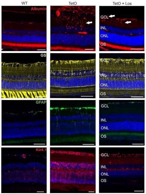
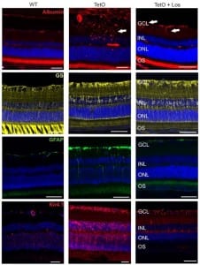
Diabetes was induced by tetracycline-inducible small hairpin RNA (shRNA) knock-down of the insulin receptor in rats (TetO). Systemic treatment consisted of an AT1-antagonist (ARB) at the onset of diabetes. 4-5 weeks later, the retina was analyzed in vivo and ex vivo. Retinal function was assessed by Ganzfeld ERG.

Retinal vessels in TetO showed calibre differences together with gliosis. The total number as well as the proportion of activated mononuclear phagocytes was increased. TetO presented loss of retinal ganglion cells (RGC). ERG indicated photoreceptor malfunction. Both the inner and outer blood-retina-barrier were affected.

The ARB-treated group presented reduced gliosis and an overall amelioration of the retinal function together with RGC recovery. No statistically significant effects on vascular and inflammatory features were found.

TetO rat represents a promising translational model for early neurovascular changes of type 2 diabetic retinopathy. ARB treatment revealed an effect on the neuronal component but not on the vasculature.
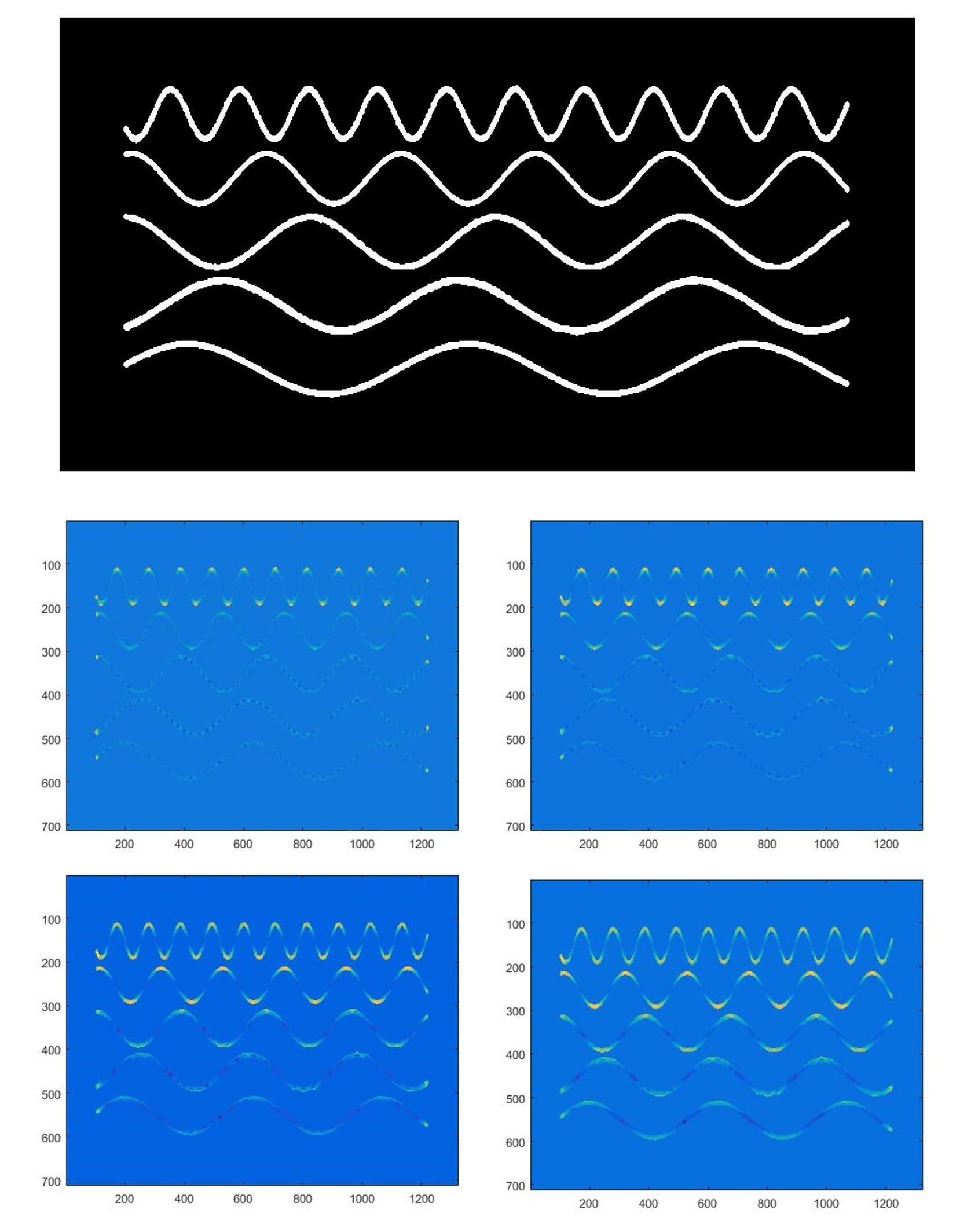
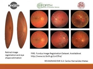
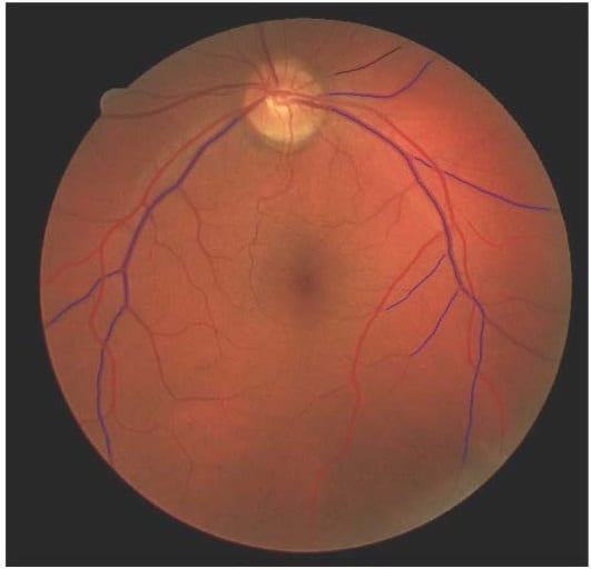
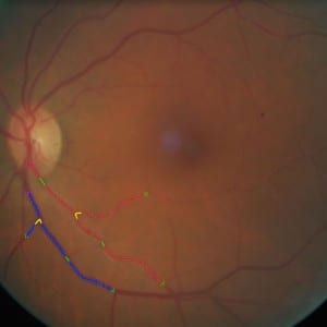
Giovanni Ometto – Progress Report 5

A software tool was created for the manual supervision of lesions resulting from the automatic algorithm. The manually supervised detection of microaneurysms in the prospective database was concluded. The new method for the quick detection of the optic disk was improved, completed with an approximate segmentation and optimised for speed and accuracy. The method for the fully automatic detection of 5 clinically relevant regions was integrated with the new, fast algorithm for the optic disk detection. A new study showed that the distribution of microaneurysms in high-risk patients was higher in the upper than in the lower hemiretina (see Figure: Cumulative distribution of microaneurysms in the left and right eye).

Giovanni Progress Report 5 - Image

Francesco Caliva – Progress Report 5

Progress Report 5 July 2016

In this reporting period, Francesco designed and implemented an algorithm that classified, as artery and vein, blood vessels along the two main retinal arcades. This study was submitted and accepted at the ARVO annual meeting.

He also collaborated with ESR 3.3, Giovanni Ometto, in a fruitful collaboration that led to the submission of a conference paper to the IEEE SPIE Medical Imaging conference.